切换至 "中华医学电子期刊资源库"

第五届中国出版政府奖音像电子网络出版物奖提名奖

中国科技核心期刊

中国科学引文数据库(CSCD)来源期刊

综述

单细胞测序技术在脓毒症免疫研究中的应用进展

  • 苏生林 1 ,
  • 马金兰 2 ,
  • 于弘明 1 ,
  • 杨晓军 , 3,
展开
  • 1.750004 银川,宁夏医科大学第一临床医学院
  • 2.750004 银川,宁夏医科大学总医院重症医学科
  • 3.750004 银川,宁夏医科大学第一临床医学院;750004 银川,宁夏医科大学总医院重症医学科
通信作者:杨晓军,Email:

Copy editor: 卫轲

收稿日期: 2024-04-30

  网络出版日期: 2024-09-06

基金资助

宁夏回族自治区重点研发计划项目(2023BEG02024)

版权

未经授权,不得转载、摘编本刊文章,不得使用本刊的版式设计,除非特别声明,本刊刊出的所有文章不代表中华医学会和本刊编委会的观点。本刊为电子期刊,以网刊形式出版。

Application of single cell sequencing technology in the study of sepsis immunity

  • Shenglin Su 1 ,
  • Jinlan Ma 2 ,
  • Hongming Yu 1 ,
  • Xiaojun Yang , 3,
Expand
  • 1.The First Clinical Medical School of Ningxia Medical University, Yinchuan 750004, China
  • 2.Department of Critical Care Medicine, General Hospital of Ningxia Medical University, Yinchuan 750004, China
  • 3.The First Clinical Medical School of Ningxia Medical University, Yinchuan 750004, China; Department of Critical Care Medicine, General Hospital of Ningxia Medical University, Yinchuan 750004, China
Corresponding author: Yang Xiaojun, Email:

Received date: 2024-04-30

  Online published: 2024-09-06

Copyright

Copyright by Chinese Medical Association No content published by the journals of Chinese Medical Association may be reproduced or abridged without authorization. Please do not use or copy the layout and design of the journals without permission. All articles published represent the opinions of the authors, and do not reflect the official policy of the Chinese Medical Association or the Editorial Board, unless this is clearly specified.

摘要

脓毒症(sepsis)是一种高病死率的临床综合征,尽管多学科方法不断发展和进步,脓毒症仍可发展为致命的器官衰竭。近年来单细胞测序技术的飞速发展,为脓毒症病理生理机制的探索提供了全新的技术支持,尤其在免疫学方面的应用使得脓毒症的精准防治成为可能。本综述通过回顾近年的相关研究,阐述脓毒症及脓毒症免疫抑制的发生发展,介绍单细胞测序技术在脓毒症及脓毒症免疫抑制研究中的应用进展,旨在探索未来使用单细胞测序技术在脓毒症精准化治疗方面应用的可能性。

本文引用格式

苏生林 , 马金兰 , 于弘明 , 杨晓军 . 单细胞测序技术在脓毒症免疫研究中的应用进展[J]. 中华重症医学电子杂志, 2024 , 10(03) : 279 -286 . DOI: 10.3877/cma.j.issn.2096-1537.2024.03.012

Abstract

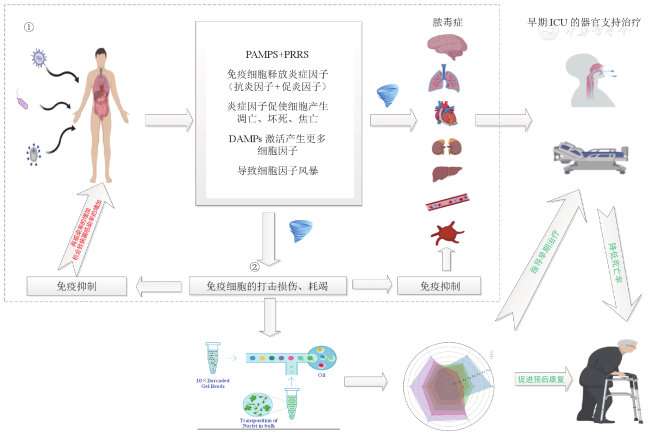
Sepsis is a clinical syndrome with a high mortality rate, and despite the development and advancement of multidisciplinary approaches, sepsis can progress to fatal organ failure. In recent years, the rapid development of single-cell sequencing technology has provided a new technical support for the exploration of the pathophysiological mechanism of sepsis, especially the application in immunology has made the precise prevention and treatment of sepsis possible. This review reviewed relevant studies in recent years, described the occurrence and development of sepsis and sepsis-induced immunosuppression, introduced the application progress of single cell sequencing technology in sepsis and sepsis-induced immunosuppression research, and aimed to explore the possibility of future application of single cell sequencing technology in the precision treatment of sepsis.

1
Singer M, Deutschman CS, Seymour CW, et al. The Third International Consensus Definitions for Sepsis and Septic Shock (Sepsis-3) [J]. JAMA, 2016, 315(8): 801-810.

2
Xie J, Wang H, Kang Y, et al. The epidemiology of sepsis in Chinese ICUs: a national cross-sectional survey [J]. Crit Care Med, 2020, 48(3): e209-e218.

3
Liu YC, Shou ST, Chai YF. Immune checkpoints in sepsis: new hopes and challenges [J]. Int Rev Immunol, 2022, 41(2): 207-216.

4
Shankar-Hari M, Calandra T, Soares MP, et al. Reframing sepsis immunobiology for translation: towards informative subtyping and targeted immunomodulatory therapies [J]. Lancet Respir Med, 2024, 12(4): 323-336.

5
Demerle KM, Angus DC, Baillie JK, et al. Sepsis subclasses: a framework for development and interpretation [J]. Crit Care Med, 2021, 49(5): 748-759.

6
Delano MJ, Ward PA. Sepsis-induced immune dysfunction: can immune therapies reduce mortality? [J]. J Clin Invest, 2016, 126(1): 23-31.

7
Seymour CW, Kennedy JN, Wang S, et al. Derivation, validation, and potential treatment implications of novel clinical phenotypes for sepsis [J]. JAMA, 2019, 321(20): 2003-2017.

8
Balch JA, Chen UI, Liesenfeld O, et al. Defining critical illness using immunological endotypes in patients with and without sepsis: a cohort study [J]. Crit Care, 2023, 27(1): 292.

9
Mchugh L, Seldon TA, Brandon RA, et al. A molecular host response assay to discriminate between sepsis and infection-negative systemic inflammation in critically ill patients: discovery and validation in independent cohorts [J]. PLoS Med, 2015, 12(12): e1001916.

10
Sweeney TE, Shidham A, Wong HR, et al. A comprehensive time-course-based multicohort analysis of sepsis and sterile inflammation reveals a robust diagnostic gene set [J]. Sci Transl Med, 2015, 7(287): 287ra71.

11
Scicluna BP, Klein Klouwenberg PM, Van Vught LA, et al. A molecular biomarker to diagnose community-acquired pneumonia on intensive care unit admission [J]. Am J Respir Crit Care Med, 2015, 192(7): 826-835.

12
Baghela A, Pena OM, Lee AH, et al. Predicting sepsis severity at first clinical presentation: The role of endotypes and mechanistic signatures [J]. EBioMedicine, 2022, 75: 103776.

13
Sweeney TE, Khatri P. Benchmarking sepsis gene expression diagnostics using public data [J]. Crit Care Med, 2017, 45(1): 1-10.

14
Sun P, Cui M, Jing J, et al. Deciphering the molecular and cellular atlas of immune cells in septic patients with different bacterial infections [J]. J Transl Med, 2023, 21(1): 777.

15
Kwok AJ, Allcock A, Ferreira RC, et al. Neutrophils and emergency granulopoiesis drive immune suppression and an extreme response endotype during sepsis [J]. Nat Immunol, 2023, 24(5): 767-779.

16
Reyes M, Filbin MR, Bhattacharyya RP, et al. An immune-cell signature of bacterial sepsis [J]. Nat Med, 2020, 26(3): 333-340.

17
Dai W, Zheng P, Wu J, et al. Integrated analysis of single-cell RNA-seq and chipset data unravels PANoptosis-related genes in sepsis [J]. Front Immunol, 2023, 14: 1247131.

18
Takeuchi O, Akira S. Pattern recognition receptors and inflammation [J]. Cell, 2010, 140(6): 805-820.

19
Deutschman CS, Tracey KJ. Sepsis: current dogma and new perspectives [J]. Immunity, 2014, 40(4): 463-475.

20
Boomer JS, To K, Chang KC, et al. Immunosuppression in patients who die of sepsis and multiple organ failure [J]. JAMA, 2011, 306(23): 2594-2605.

21
Joshi I, Carney WP, Rock EP. Utility of monocyte HLA-DR and rationale for therapeutic GM-CSF in sepsis immunoparalysis [J]. Front Immunol, 2023, 14: 1130214.

22
Leijte GP, Rimmelé T, Kox M, et al. Monocytic HLA-DR expression kinetics in septic shock patients with different pathogens, sites of infection and adverse outcomes [J]. Crit Care, 2020, 24(1): 110.

23
Asmussen A, Busch HJ, Helbing T, et al. Monocyte subset distribution and surface expression of HLA-DR and CD14 in patients after cardiopulmonary resuscitation [J]. Sci Rep, 2021, 11(1): 12403.

24
De Roquetaillade C, Dupuis C, Faivre V, et al. Monitoring of circulating monocyte HLA-DR expression in a large cohort of intensive care patients: relation with secondary infections [J]. Ann Intensive Care, 2022, 12(1): 39.

25
Boeddha NP, Kerklaan D, Dunbar A, et al. HLA-DR expression on monocyte subsets in critically ill children [J]. Pediatr Infect Dis J, 2018, 37(10): 1034-1040.

26
Gouel-Chéron A, Allaouchiche B, Guignant C, et al. Early interleukin-6 and slope of monocyte human leukocyte antigen-DR: a powerful association to predict the development of sepsis after major trauma [J]. PLoS One, 2012, 7(3): e33095.

27
Cour-Andlauer F, Morrow BM, Mcculloch M, et al. Decreased human leukocyte antigen DR on circulating monocytes expression after severe pediatric trauma: an exploratory report [J]. Pediatr Crit Care Med, 2021, 22(5): e314-e323.

28
Wu JF, Ma J, Chen J, et al. Changes of monocyte human leukocyte antigen-DR expression as a reliable predictor of mortality in severe sepsis [J]. Crit Care, 2011, 15(5): R220.

29
Remy S, Kolev-Descamps K, Gossez M, et al. Occurrence of marked sepsis-induced immunosuppression in pediatric septic shock: a pilot study [J]. Ann Intensive Care, 2018, 8(1): 36.

30
Winkler MS, Rissiek A, Priefler M, et al. Human leucocyte antigen (HLA-DR) gene expression is reduced in sepsis and correlates with impaired TNFα response: a diagnostic tool for immunosuppression? [J]. PLoS One, 2017, 12(8): e0182427.

31
Pei F, Zhang GR, Zhou LX, et al. Early immunoparalysis was associated with poor prognosis in elderly patients with sepsis: secondary analysis of the ETASS study [J]. Infect Drug Resist, 2020, 13: 2053-2061.

32
Kox M, Frenzel T, Schouten J, et al. COVID-19 patients exhibit less pronounced immune suppression compared with bacterial septic shock patients [J]. Crit Care, 2020, 24(1): 263.

33
Yi JS, Cox MA, Zajac AJ. T-cell exhaustion: characteristics, causes and conversion [J]. Immunology, 2010, 129(4): 474-481.

34
Pfortmueller CA, Meisel C, Fux M, et al. Assessment of immune organ dysfunction in critical illness: utility of innate immune response markers [J]. Intensive Care Med Exp, 2017, 5(1): 49.

35
Stubbington MJT, Rozenblatt-Rosen O, Regev A, et al. Single-cell transcriptomics to explore the immune system in health and disease [J]. Science, 2017, 358(6359): 58-63.

36
Chen H, Ye F, Guo G. Revolutionizing immunology with single-cell RNA sequencing [J]. Cell Mol Immunol, 2019, 16(3): 242-249.

37
Stoeckius M, Hafemeister C, Stephenson W, et al. Simultaneous epitope and transcriptome measurement in single cells [J]. Nat Methods, 2017, 14(9): 865-868.

38
Peterson VM, Zhang KX, Kumar N, et al. Multiplexed quantification of proteins and transcripts in single cells [J]. Nat Biotechnol, 2017, 35(10): 936-939.

39
Dey SS, Kester L, Spanjaard B, et al. Integrated genome and transcriptome sequencing of the same cell [J]. Nat Biotechnol, 2015, 33(3): 285-289.

40
Macaulay IC, Haerty W, Kumar P, et al. G&T-seq: parallel sequencing of single-cell genomes and transcriptomes [J]. Nat Methods, 2015, 12(6): 519-522.

41
Bian S, Hou Y, Zhou X, et al. Single-cell multiomics sequencing and analyses of human colorectal cancer [J]. Science, 2018, 362(6418): 1060-1063.

42
Hu Y, Huang K, An Q, et al. Simultaneous profiling of transcriptome and DNA methylome from a single cell [J]. Genome Biol, 2016, 17: 88.

43
Wang X, Allen WE, Wright MA, et al. Three-dimensional intact-tissue sequencing of single-cell transcriptional states [J]. Science, 2018, 361(6400): eaat5691.

44
Peng G, Suo S, Chen J, et al. Spatial transcriptome for the molecular annotation of lineage fates and cell identity in mid-gastrula mouse embryo [J]. Dev Cell, 2016, 36(6): 681-697.

45
Chen J, Suo S, Tam PP, et al. Spatial transcriptomic analysis of cryosectioned tissue samples with Geo-seq [J]. Nat Protoc, 2017, 12(3): 566-580.

46
Pei F, Yao RQ, Ren C, et al. Expert consensus on the monitoring and treatment of sepsis-induced immunosuppression [J]. Mil Med Res, 2022, 9(1): 74.

47
Inoue S, Suzuki-Utsunomiya K, Okada Y, et al. Reduction of immunocompetent T cells followed by prolonged lymphopenia in severe sepsis in the elderly [J]. Crit Care Med, 2013, 41(3): 810-819.

48
Wang T, Zhang X, Liu Z, et al. Single-cell RNA sequencing reveals the sustained immune cell dysfunction in the pathogenesis of sepsis secondary to bacterial pneumonia [J]. Genomics, 2021, 113(3): 1219-1233.

49
Misheva M, Kotzamanis K, Davies LC, et al. Oxylipin metabolism is controlled by mitochondrial β-oxidation during bacterial inflammation [J]. Nat Commun, 2022, 13(1): 139.

50
Wu M, Huang Z, Huang W, et al. microRNA-124-3p attenuates myocardial injury in sepsis via modulating SP1/HDAC4/HIF-1α axis [J]. Cell Death Discov, 2022, 8(1): 40.

51
Almansa R, Heredia-Rodríguez M, Gomez-Sanchez E, et al. Transcriptomic correlates of organ failure extent in sepsis [J]. J Infect, 2015, 70(5): 445-456.

52
Xie X, Le L, Fan Y, et al. Autophagy is induced through the ROS-TP53-DRAM1 pathway in response to mitochondrial protein synthesis inhibition [J]. Autophagy, 2012, 8(7): 1071-1084.

53
Meydan S, Marks J, Klepacki D, et al. Retapamulin-assisted ribosome profiling reveals the alternative bacterial proteome [J]. Mol Cell, 2019, 74(3): 481-493.e496.

54
Zheng W, Wang X, Liu J, et al. Single-cell analyses highlight the proinflammatory contribution of C1q-high monocytes to Behçet’s disease [J]. Proc Natl Acad Sci U S A, 2022, 119(26): e2204289119.

55
Murao A, Jha A, Aziz M, et al. Transcriptomic profiling of immune cells in murine polymicrobial sepsis [J]. Front Immunol, 2024, 15: 1347453.

56
He XL, Chen JY, Feng YL, et al. Single-cell RNA sequencing deciphers the mechanism of sepsis-induced liver injury and the therapeutic effects of artesunate [J]. Acta Pharmacol Sin, 2023, 44(9): 1801-1814.

57
Gu J, Xu Y, Hua D, et al. Role of artesunate in autoimmune diseases and signaling pathways [J]. Immunotherapy, 2023, 15(14): 1183-1193.

58
Chen G, Ren C, Xiao Y, et al. Time-resolved single-cell transcriptomics reveals the landscape and dynamics of hepatic cells in sepsis-induced acute liver dysfunction [J]. JHEP Rep, 2023, 5(6): 100718.

59
Zhang YF, Yi ZJ, Zhang WF, et al. Single-cell sequencing reveals MYOF-enriched monocyte/macrophage subcluster as a favorable prognostic factor in sepsis [J]. Adv Biol (Weinh), 2024: e2300673.

60
Li R, Li X, Zhao J, et al. Mitochondrial STAT3 exacerbates LPS-induced sepsis by driving CPT1a-mediated fatty acid oxidation [J]. Theranostics, 2022, 12(2): 976-998.

61
Wen M, Cai G, Ye J, et al. Single-cell transcriptomics reveals the alteration of peripheral blood mononuclear cells driven by sepsis [J]. Ann Transl Med, 2020, 8(4): 125.

62
She H, Tan L, Wang Y, et al. Integrative single-cell RNA sequencing and metabolomics decipher the imbalanced lipid-metabolism in maladaptive immune responses during sepsis [J]. Front Immunol, 2023, 14: 1181697.

63
Villani AC, Satija R, Reynolds G, et al. Single-cell RNA-seq reveals new types of human blood dendritic cells, monocytes, and progenitors [J]. Science, 2017, 356(6335): eaah4573.

64
Ning J, Fan X, Sun K, et al. Single-cell sequence analysis combined with multiple machine learning to identify markers in sepsis patients: LILRA5 [J]. Inflammation, 2023, 46(4): 1236-1254.

65
Franzén O, Gan LM, Björkegren JLM. PanglaoDB: a web server for exploration of mouse and human single-cell RNA sequencing data [J]. Database (Oxford), 2019: baz046.

66
Cho DS, Schmitt RE, Dasgupta A, et al. Single-cell deconstruction of post-sepsis skeletal muscle and adipose tissue microenvironments [J]. J Cachexia Sarcopenia Muscle, 2020, 11(5): 1351-1363.

文章导航

/